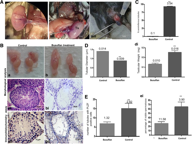
Fig. 4.
a–e Effect of treatment with busulfan in mouse testes after 6 weeks, before cSSC xenotransplantation. a Technique for the transplantation of cSSCs into the mouse testes. ai–aiii Insulin needle inserted into the efferent duct (asterisk) located the testicular artery cranially (arrow) between the tail of the epididymis (Ep) and testis (Ts) to help delineate the ducts and the testicular network, and then, all seminiferous tubules were filled with a small amount of blue dye solution (Trypan Blue). b Macroscopic and microscopic analyses of the mouse testes before xenotransplantation: control and treated (busulfan) groups (scale bar = 1 cm, 20 μm). ai–aii Control group, spermatogenesis was normal. It was possible to observe complete spermatogenesis (spermatogonia, spermatocytes, and spermatozoa) in the mouse seminiferous tubules. b–bii Treated group did not exhibit germ cells in the germinative epithelium. In the seminiferous tubules, only Sertoli cells (Se) were present. c Graphic showing the statistical analysis of the mean number of seminiferous tubules exhibiting normal spermatogenesis in the control and treated groups. d Graphic showing the changes in tubular diameter and testicular weight in the control and treatment groups. e Graphic showing the number of cells that expressed the PLZF marker. ei Number of spermatozoa after busulfan treatment was performed to confirm busulfan efficiency. **Significant difference (p < 0.05). (Scale bar = 20 μm)

