Fig. 2.
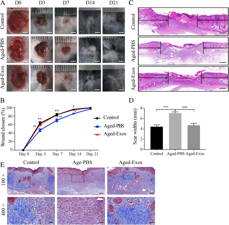
ESC-Exos promoted pressure ulcer wound healing in aged mice. a Gross view of wounds treated with ESC-Exos or PBS in aging mice and wounds with PBS in the young control group, at days 3, 7, 14, and 21 post-wounding. Scale bar, 2 mm. b The rate of wound closure of three groups. n = 6 per group. **P < 0.01; *P < 0.05 Aged-PBS versus control group; ##P < 0.01; #P < 0.05 Aged-Exos versus Aged-PBS group. c H&E staining of wound sections from three groups at 7 days after initial treatment. The black arrows indicate the edges of the scar. n = 3 per group. Scale bar, 500 μm. d Quantification of the scar widths. n = 3 per group. ***P < 0.001 Aged-PBS versus control group; ###P < 0.001 Aged-Exos versus Aged-PBS group. e Masson’s trichrome staining of wound sections from three groups. n = 3 per group. Scale bar, 100 μm (top) or 25 μm (bottom)

