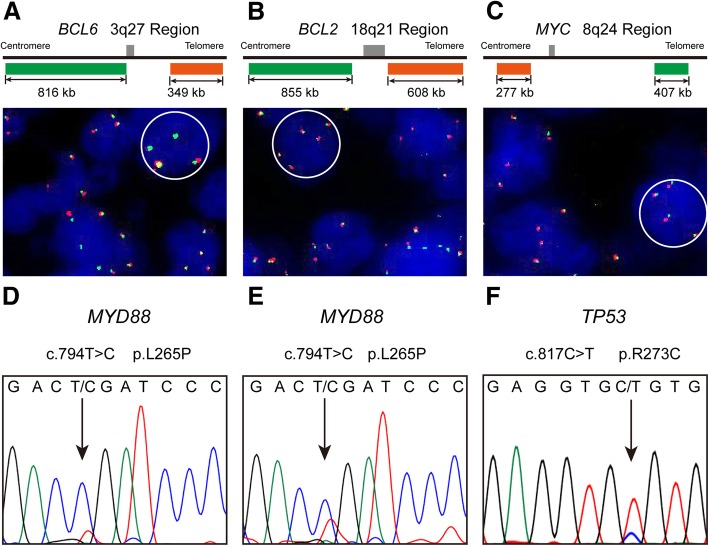
Fig. 2.
Representative results of fluorescence in situ hybridization (FISH) analysis and Sanger sequencing of primary CNS A-DLBCL. The FISH analysis for patient 1 showed extra copy signals for BCL6 (a), BCL2 (b) and MYC (c). MYD88 L265P mutations in patients 1 (d) and 2 (e), in which a CTG (leucine) codon was changed to a CCG (proline) codon. TP53 R273C mutation of patient 3 (f), in which a CGT (arginine) codon was changed to a TGT (cysteine) codon

