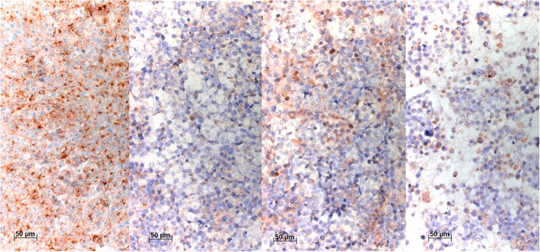
Fig. 3.
Immunocytochemical stainings of NCI-H69s with VenA, AbcA, NovA, and TherA (left to right). VenA shows an intense membranous but also a cytoplasmic staining, the latter concentrated as dots, most likely representing accumulation within endoplasmic reticulum. AbcA stained less than half of the cells, whereas NovA and TherA stained approximately 50% of the cells, but in variable intensity. Bar 50 μm

