. 2019 Feb;16(1):55–70. doi: 10.20892/j.issn.2095-3941.2018.0157

2.

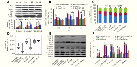
(A) Western blot analysis of the effects of insulin and estradiol treatment on the phosphorylation of ER, InsR, and IRS-1 in Ishikawa cells. (B) BrdU ELISA of the effects of estradiol stimulation on Ishikawa cell proliferation after knockdown of ER and/or InsR. (C) Flow cytometry analysis of the effects of estradiol stimulation on Ishikawa cell cycle distribution after knockdown of ER and/or InsR. (D) The percentage of apoptotic Ishikawa cells (after knockdown of ER and/or InsR) stimulated with estradiol was detected via flow cytometry analysis using Annexin V-FITC antibodies. (E, F) Western blot analysis of the effects of estradiol treatment on the ratios of phosphorylated (p)-InsR/InsR, p-IRS-1/IRS-1, p-Akt/Akt, p-MAPK/MAPK, and p-ERK/ERK in Ishikawa cells after knockdown of ER and/or InsR. *<italic>P</italic> < 0.05 for all experiments.

Estrogen promotes EC cell progression via InsR by activating the PI3K and MAPK pathways.