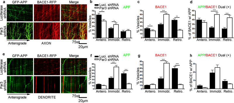
Figure 4. Loss of Par3 increases APP and BACE1 convergence in axons.
(a) Hippocampal neurons were transfected with indicated constructs together with GFP-APP and hBACE1-RFP. Live images were captured on DIV11 and kymographs were generated to trace APP and hBACE1 trafficking in axons. (b) Quantification of the mobility of APP containing vesicles in axons (anterograde, immobile and retrograde). (c) Quantifications of the mobility of BACE1 containing vesicles in axons. (d) Quantifications of the percentage of BACE1 vesicles that are co-trafficking with APP vesicles in axons. N (cells) = 17 (Luciferase shRNA), 14 (Par3 shRNA) from 6 independent experiments, with 20–50 vesicles analyzed per movie. (e) Hippocampal neurons were transfected with indicated constructs together with GFP-APP and hBACE1-RFP. Live images were captured on DIV11 and kymographs were generated to trace APP and hBACE1 trafficking in dendrites. (f) Quantification of the mobility of APP containing vesicles in dendrites (anterograde, immobile and retrograde). (g) Quantifications of the mobility of BACE1 containing vesicles in dendrites. (h) Quantifications of the percentage of BACE1 vesicles that are co-trafficking with APP vesicles in dendrites. N (cells) = 14 (Luciferase shRNA), 16 (Par3 shRNA) from 4 independent experiments, with 10–30 vesicles analyzed per movie. Data were expressed as Mean ± SEM with Student’s t test: *p<0.05; ** p<0.01; *** p<0.001. Scale bar: 75s/20μm.

