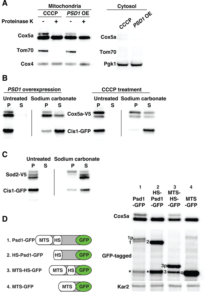
Fig. 2. Mitochondrial precursors accumulate on the surface of the organelle and in the translocase during import stress.
(A) Mitochondria were isolated by differential centrifugation from cells treated with 20μM CCCP for 1h or cells overexpressing PSD1 for 6h. Cox5a-V5, Tom70-mCherry and Cox4 or Pgk1 were detected in mitochondria and cytosol fractions. Mitochondria were treated with 50μg/ml proteinase K. Tom70 served as an outer membrane control protein; Cox4 as a matrix control protein. OE, overexpression. (B) Mitochondria were isolated from cells expressing COX5a-V5 and CIS1-GFP and overexpressing PSD1 for 6h or cells treated with 20μM CCCP for 1h. Sodium carbonate treated or untreated mitochondria were centrifuged to separate insoluble proteins [pellet (P)] from soluble proteins [supernatant (S)]. Samples were analyzed by immuno-blot analysis. Cis1-GFP served as a peripheral outer membrane protein control. (C) Mitochondria were isolated from cells overexpressing PSD1 for 6h. Mitochondria were treated as in (B) to analyze Sod2-V5 by immuno-blot analysis. (D) Left panel: PSD1-GFP constructs used in the analysis. MTS, mitochondrial targeting sequence; HS, hydrophobic segment. Right panel: immuno-blot blot of Cox5a-V5 and Psd1-GFP fusion proteins following overexpression of PSD1-GFP fusion genes for 4h. Kar2 was used as a loading control. Numbering on the immuno-blot indicates the mature form of the GFP-tagged proteins. The letter p following this number identifies the precursor form of proteins. Asterisks identify a proteolytic cleavage product of Psd1 known as the α subunit (51).

