
Fig. 4.
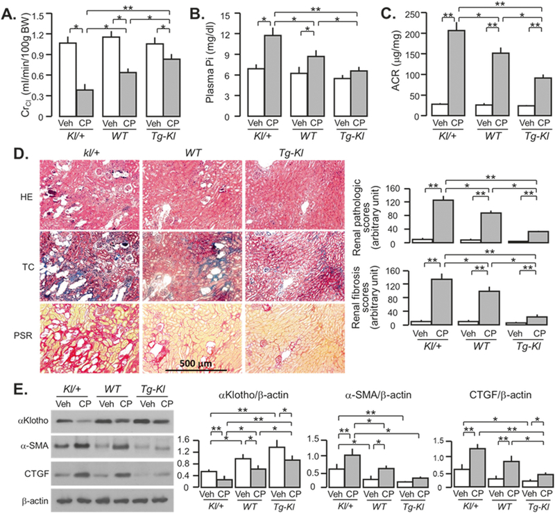
αKlotho-deficient mice progress more rapidly to CKD after cisplatin (CP) injection. Mice with three different levels of αKlotho (low kl/+; normal WT; and high Tg-Kl) at 3 months old were intraperitoneally injected with normal saline as vehicle or CP (10mg/Kg), and fed high phosphate (Pi) rodent chow 2 weeks after the injection for 18 weeks. a Creatinine clearance (ClCr). b Plasma phosphate (Pi). c Albumin-to-creatinine ratio (ACR). d Kidney histology assessed by HE, TC, and PSR stain (scale bar = 500 μm); and semi-quantitative assessment (right panel including chronic pathologic score based on HE stain and renal fibrosis score based on PSR stain). e αKlotho and fibrotic markers in the kidney. Left panel: representative immune-blotting for αKlotho, α-SMA, and CTGF protein in total kidney lysates. Right panel: summary of immunoblots in arbitrary units from all immunoblots. Data are expressed as means ± SD from each group and statistical significance was evaluated by one-way ANOVA followed by Student–Newman–Keuls post hoc test, and significance was accepted when *P < 0.05; **P < 0.01 between two groups. HE Hematoxylin and Eosin stain; TC Trichrome stain; PSR Picrosirius red stain