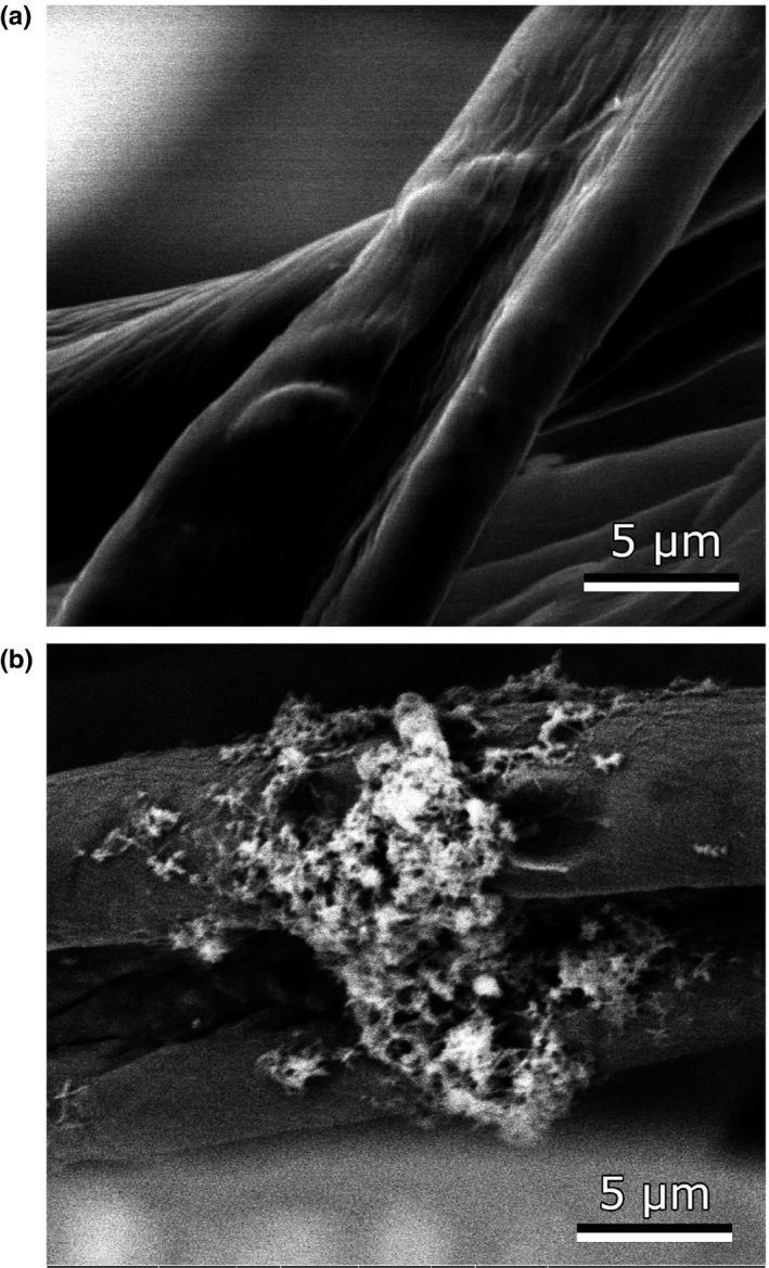
Figure 3.

Scanning electron microscope (SEM) images of un‐inoculated cotton balls (a) and cotton balls cultured in the presence of P. luteoviolacea 2ta16. In this image, a microcolony of bacterial cells can be seen adhering to the surface of two cotton fibers
