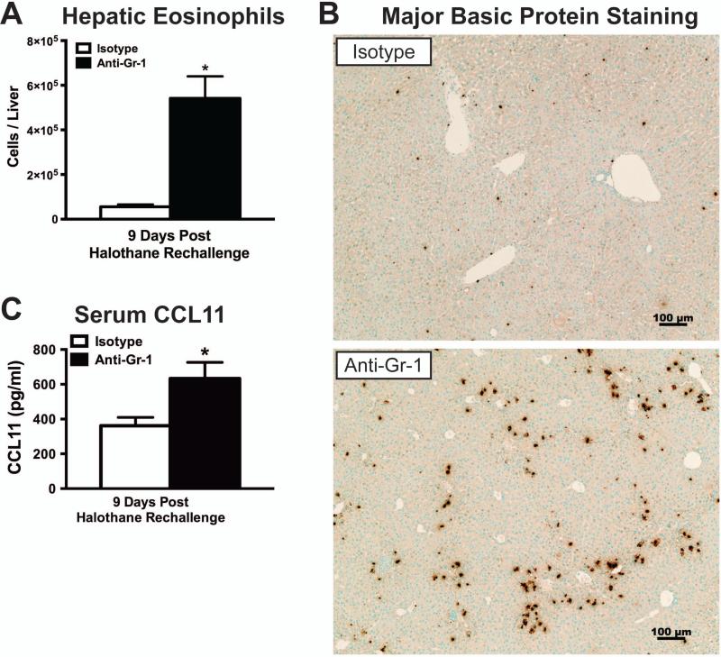
Figure 4. Depletion of hepatic MDSCs prior to initial halothane treatment resulted in elevated hepatic eosinophil infiltration.
(A) Hepatic eosinophils (SSChigh CD11c− Gr-1low Siglec-Fhigh) were measured by flow cytometry 9 days after halothane rechallenge with N=14 per group from 2 independent experiments. (B) Representative photomicrographs of Major Basic Protein (MBP) staining for the detection of eosinophils in isotype or anti-Gr-1 treated mice 9 days after rechallenge with halothane. (C) Serum protein concentrations of CCL11 were measured in isotype or anti-Gr-1 treated mice 9 days after halothane rechallenge with N=11 per group from 3 independent experiments. All data reported as means ± SEM. *P<0.05 versus isotype-treated group.

