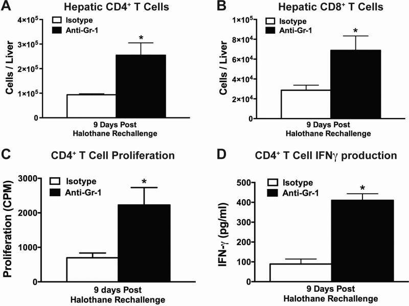
Figure 5. Depletion of hepatic MDSCs prior to initial halothane treatment resulted in increased number of hepatic CD4+ and CD8+ T cells after halothane rechallenge.
Total number of hepatic CD4+ (A) or CD8+ (B) T cells per liver from isotype or anti-Gr-1 treated mice were quantified at 9 days post halothane rechallenge. (C) Hepatic CD4+ T cells isolated 9 days post halothane rechallenge from either isotype or anti-Gr-1 treated mice were cultured for 96 hours in the presence of microsomal protein extract from halothane or oil-treated mice along with naïve irradiated mouse splenocytes as antigen presenting cells. Proliferation was measured by [3H]-thymidine incorporation during the last 24 hours of incubation. The results were corrected by subtracting the counts per minute (CPM) from the wells incubated with microsomal extracts from oil treated mice. (D) IFNγ protein levels in cell-culture supernatant were assessed in CD4+ T cells isolated, from isotype or anti-Gr-1 treated mice, 9 days post halothane rechallenge. Cells were cultured for 72 hours in the presence of microsomal protein extract from halothane or oil treated mice along with naïve irradiated mouse splenocytes as antigen presenting cells. All data reported as mean ± SEM of N=4-6 mice per group. *P<0.05 versus isotype-treated group.

