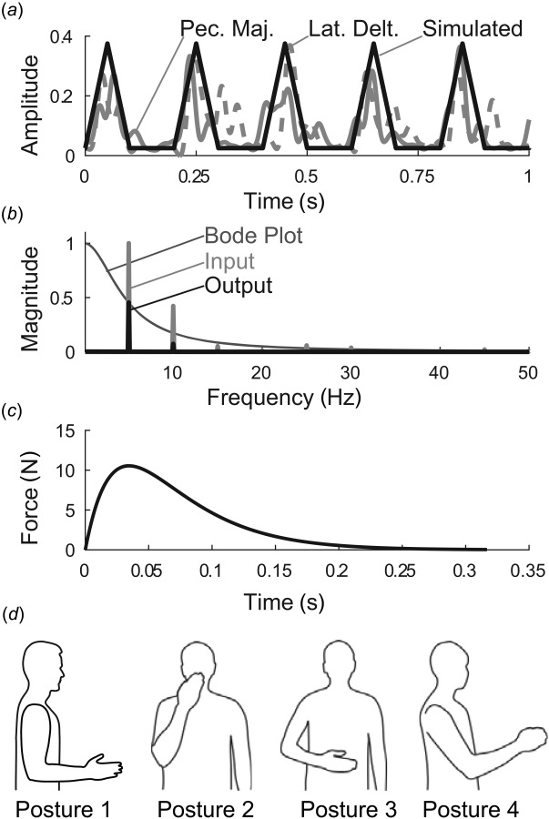
Fig. 2.

Methodological details. (a) input muscle activity was approximated by triangular waves, based on experimentally observed sEMG in tremor patients. Shown are detrended, rectified, and low-pass filtered sEMG signals from pectoralis major (solid gray) and lateral deltoid (dashed gray) muscles from a subject with severe tremor, compared to triangular waves (black), (b) Magnitude ratio of first submodel (excitation–contraction dynamics, with default values and ), along with the Fourier transforms of the input signal, (5 Hz triangle wave of width 110 ms), and the output, , (c) The dynamics of the first submodel (muscle excitation–contraction dynamics) are illustrated by the submodel's impulse response, which represents a muscle twitch (simulated using default values and ), (d) Postures included in our simulation. Posture 1 is the default posture. Postures 2–4 were used in the sensitivity analysis. Posture 2: hand in front of mouth, representing feeding and grooming activities; Posture 3: hand in workspace in front of abdomen, representing many activities of daily living; and Posture 4: arm somewhat outstretched, representing reaching. Joint angles for each posture are given in Ref. [25].
