Fig. 2.
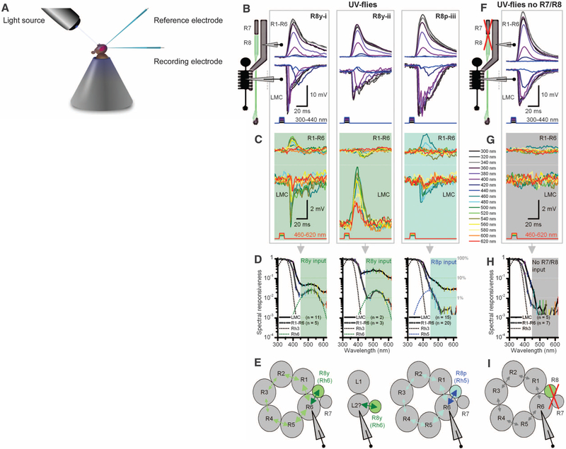
Motion pathway receives information from R8 photo-receptors in color pathway. (A) Schematic of in vivo recordings using monochromatic light. (B) Three intracellular response types from R1–R6 photoreceptors and postsynaptic LMCs to subsaturating short-wavelength (300 to 420 nm) pulses of equal energy. (C) Stimulating the same representative cells (B, i to iii) with equal long-wavelength (420 to 620 nm) pulses, which are undetectable by Rh3, reveals unexpected responses both in polarity and spectral responsiveness. (D) Spectral responsiveness of R1–R6 and LMC outputs confirms a second peak or bulge, which matches the sensitivity of R8y (Rh6) or R8p (Rh5) photoreceptors; right graph also includes cells with subtle long-wavelength range extensions. (E) Results suggest a model (29) in which information spreads via gap junctions from each R8 to R6 axon, and further to other photoreceptors in the same cartridge, before transmission to LMCs. Depolarizing responses (420 to 620 nm) in some LMCs suggest gap junctions between L2 and R8 axons. (F) In UV flies in which R1–R8 phototransduction was deactivated (norpA36) and then rescued only in R1–R6 (i.e., light-insensitive R7/R8), R1–R6 and LMC have a sensitivity to short-wavelength stimuli that is similar to when R7/R8 are functional (B). (G) These R1–R6s and LMCs cannot detect 460 to 620 nm. (H) Their spectral responsiveness matches the Rh3 sensitivity of dissociated cells (Fig. 1C). (I) With light-insensitive R7/R8s, information processing occurs independently within the motion pathway.

