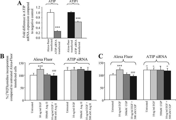
Fig. 3.
A : Fold-decreasein ATIP and ATIP1mRNA expression following transfection of ATIP siRNA into PC3 cells.*** indicates a significant decreasein ATIP mRNA expression, P < 0.001.B,C : % Change in DNA synthesis in PC3 cells transiently transfected with either ATIP siRNA or Alexa Fluor (negative control) in the presence or absence of (B) 10 ng/ml EGF and 500 nM Ang II and the combination of EGF and Ang II; or (C) 10 ng/mlEGF and 500 nM CGP42112A and the combination of EGF and CGP42112A for 72 hr.***denotes that there is a significant increase in DNA synthesis compared to untreated PC3 cells transfected with the same plasmid(P < 0.001, repeated measures ANOVA test with Tukey post-test).- denotes that there is a significant difference in DNA synthesis between ATIP silenced and untreated control cells.### denotes that there is significantly less DNA synthesisin Alexa Fluor-transfected cells treated with the combination of EGF and Ang II or EGF and CGP42112A thanin cells treated with EGF P < 0.001.Each value represents the mean + standard error of the mean of four independent experiments.

