Fig. 7.
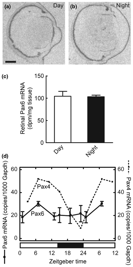
Daily expression of Pax6 in the retina of the adult rat. (a–c) Quantitative radiochemical in situ hybridization analysis of a diurnal expression of Pax6 in the retina of the adult rat housed under a 12 : 12 light/dark schedule. (a) Autoradiograph of a section of the eyeball from an animal killed at mid-day zeitgeber time (ZT6). (b) Autoradiograph of a section of the eyeball from an animal killed at midnight (ZT18). Scale bar, 1 mm. (c) Densitometric quantification of Pax6 mRNA in the rat retina. Values on the bar graph represent the mean ± SEM of four animals in each group. No day-night differences were detected (two-tailed Student’s t-test, t6 = 0.12, p = 0.91). (d) Quantitative RT-PCR analysis of daily expression of Pax6 in the retina of the adult rat housed under a 14 : 10 light/dark schedule. Values on graphs represent the mean ± SEM of three different pools of three retinae at each time point. For comparison, the daily pattern of Pax4 expression is shown (dashed line). Diurnal differences in Pax6 expression were not detected in the retina (one-way ANOVA, F5,12 = 0.66, p = 0.66).

