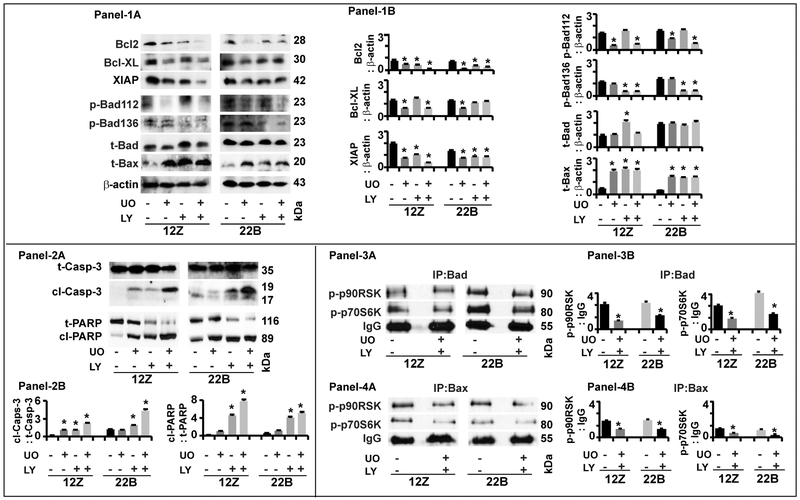
Fig-7: Effects of ERK1/2 and AKT pathways on intrinsic apoptosis pathway proteins in human endometriotic cells.
Panel-1: Antiapoptotic and proapoptotic proteins. (1A) Representative Immunoblot and (1B) Histogram. Panel-2: Caspase-3 and PARP proteins. (2A) Representative Immunoblot and (2B) Histogram. Panel-3: Interactions between Bad and p-p90RSK and p-p70S6K. (3A) Representative immunoprecipitation/immunoblot and (3B) Histogram. Panel-4: Interactions between Bax and p-p90RSK and p-p70S6K. (4A) Representative immunoprecipitation/immunoblot and (4B) Histogram. The human endometriotic epithelial cells 12Z and stromal cells 22B were treated with MEK1/2 inhibitor (U0126, 20μm) to suppress ERK1/2 pathway or PI3K inhibitor (LY294002, 50μm) to suppress AKT pathway for 24h. Expression of intrinsic apoptosis pathway proteins were analyzed by western blot. β-actin protein was measured as an internal control. Protein-protein interaction was determined by immunoprecipitation. IgG was measured as internal control. The densitometry of autoradiograms was performed using an Alpha Imager. Data expressed in integrated density value (IDV). *- control vs. treatment, p<0.05, n=3. See Materials and Method section for additional experimental details.

