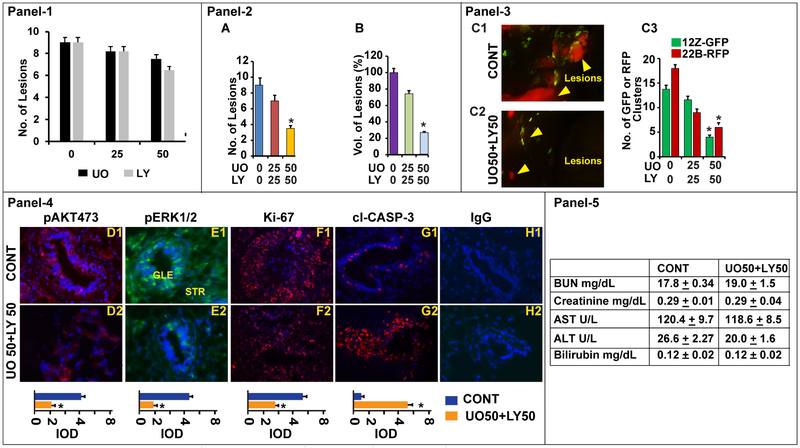
Fig-8: Effects of ERK1/2 and AKT pathways on growth and survival of endometriotic lesions.
A mixture of human endometriotic epithelial cells 12Z-GFP and stromal cells 22B-RFP suspension was injected into the peritoneal cavity of Rag2g(c) mice and peritoneal endometriosis was induced (day 1). The endometriosis mice were treated with MEK1/2 inhibitor U0126 (UO@ 0, 25, 50 mg/kg) to suppress ERK1/2 pathway and/or PI3K inhibitor LY294002 (LY@ 0, 25, 50 mg/kg) to suppress AKT pathway from days 15-28. The mice were necropsied on day 29-30 on E2-phase of the estrus cycle. Panel-1: Histogram shows the dose-dependent inhibitory effects of ERK1/2 (n=3) or AKT (n=3) pathway on growth of endometriotic lesions. Panel-2: Histogram shows the dose-dependent effects of combined inhibition of ERK1/2 or AKT pathways (n=6) on growth of endometriotic lesions, (A) number of lesions and (B) volume of lesions. Panel-3: Dose-dependent effects @ UO50/LY50 is shown. (C1-C2) Fluorescence zoomstereo microscopy examination of dissemination of 12Z-GFP and 22B-RFP cells of endometriotic lesions in the peritoneal cavity, yellow arrows show the lesions. (C3) Histogram shows number of 12Z-GFP and 22B-RFP cells in these endometriotic lesions. Panel-4: Expression of (D1-D2) pAKT, (E1-E2) pERK1/2, (F1-F2) ki-67, and (G1-G2) cl-Caspase-3 proteins in the endometriotic lesions. (H1-H2): Negative control IgG. GLE: Glandular epithelial cells. STR: Stromal cells. Relative expression was quantified using Image Pro-Plus. Panel-5: Biochemical profile. *- control vs. treatment, p<0.05, n=6 mice.

