
Figure 6.
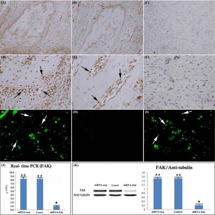
shRNA knockdown of Fak expression in the distracted callus. Immunohistochemical image showing FAK expression in the shRNA‐neg (A, D), control group (B, E) and shRNA‐Fak (C, F) (original magnification ×100 and ×400). No FAK protein expression was present with FAK antibody staining in the shRNA‐Fak group. (C, F) The infected callus region showing GFP expression in the shRNA‐neg (G), control (H) and shRNA‐Fak group (I). Mesenchymal cells in callus region were efficiently infected by the lentivirus constructs and express GFP in both shRNA‐neg (G) and shRNA‐Fak group (I). RT‐PCR and Western blot analysis showed that the level of FAK was significantly reduced in shRNA‐Fak group (J, K). (n = 8 rats/group) (## vs ★: P < .01) (n = 8 rats/group)