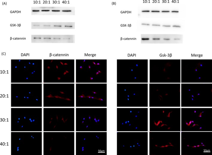
Figure 6.

A, Protein expression of β‐catenin in DPSCs plated on different elastic substrates culturing for 14 days. B, Protein expression of Gsk‐3β measured by Western blotting in DPSCs seeded on different elastic substrates culturing for 21 days. C, Immunofluorescence of β‐catenin and Gsk‐3β in DPSCs seeded on substrates with different stiffness for 7 days. β‐catenin (red), cell nuclei (blue). Scale bars are 50 μm
