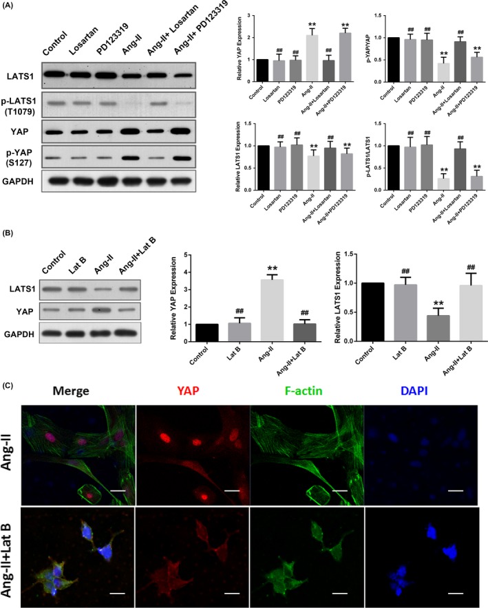
Figure 6.

Ang II regulated YAP activity via AT1R‐ and F‐actin‐dependent mechanisms. Quiescent rat VSMCs were pretreated under the following conditions: 1 μmol/L losartan for 30 min; 1 μmol/L PD 123319 for 30 min; or 500 nmol/L latrunculin B for 20 min. Then, all cells were stimulated with Ang II (10−7 mol/L for 24 h) and harvested for the assay. A and B, Densitometric quantification and representative Western blotting for YAP and LATS1 were shown. The data were normalized to GAPDH and expressed relative to the control values (set to 1). C, Representative immunofluorescence staining of YAP (red) and F‐actin (Alexa Fluor® 488 Phalloidin, green). All sections were counterstained with DAPI to visualize the nuclei (blue). Images were acquired using a Zeiss confocal microscope. Scale bars represent 10 μm. *P < 0.05 compared with the control group, **P < 0.01 compared with the control group; #P < 0.05 compared with the Ang II group, ##P < 0.01 compared with the Ang II group. Lat B, latrunculin B. GAPDH, glyceraldehyde phosphate dehydrogenase
