
Figure 1.
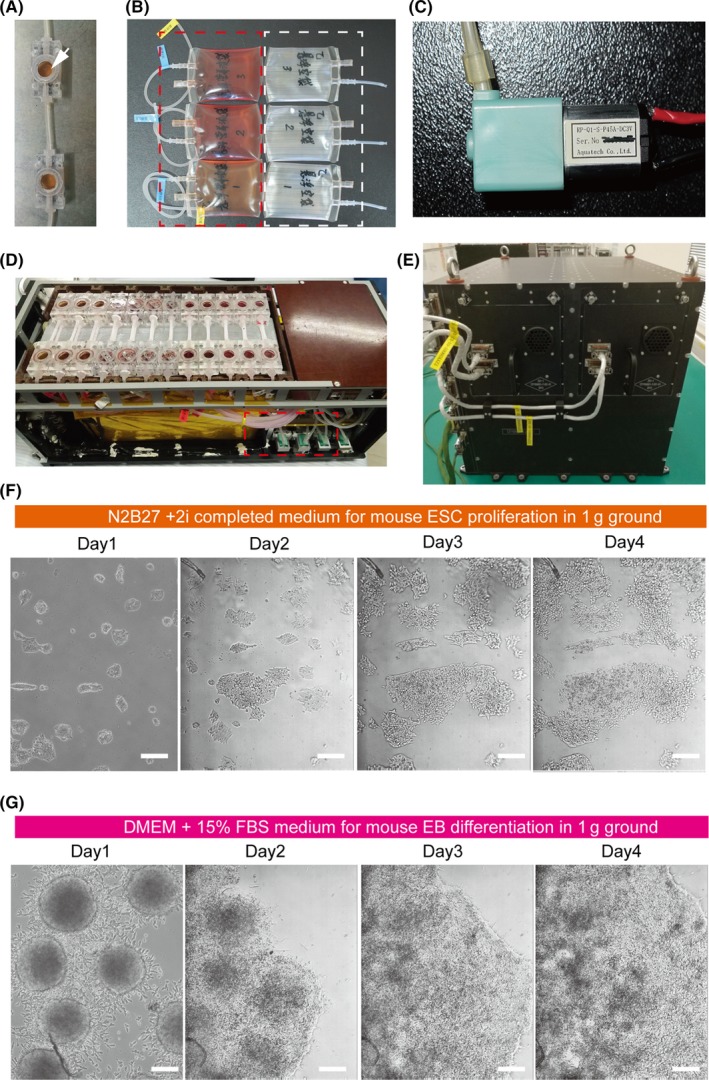
Establishment of an automated culture system to culture mouse embryonic stem cells (mESCs) and embryoid bodies (EBs). A, In our cell culture chamber, real‐time growth of cells can be monitored by microscopy through the window (white arrow). B, Pictures of the medium storage bags (red dotted line) and waste bags (white dotted line). C, Micro peristaltic pumps, which provide medium change with a flow rate of 400 μL/min in these experiments. D, Cell culture module (CCM), consisting of 2 connecting space culture chambers linked with the bags and with the pump. E, The electronic container, together with CCM, forms the complete experimental hardware, called the bioreactor. F, Representative bright field images of mESCs cultured with N2b27/2i medium using the automated culture system in ground conditions. Scale bars = 100 μm. G, Representative bright field images of EBs cultured with differentiation medium using the automated culture system in ground conditions. Scale bars = 200 μm