
Figure 5.
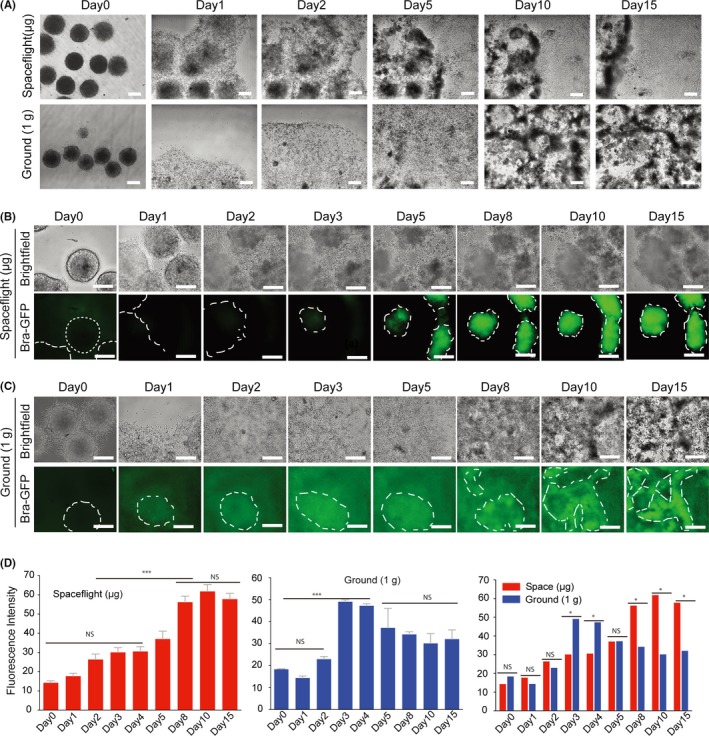
Cell adhesion, outgrowth and differentiation of Brachyury‐GFP EBs cultured in space and ground environment. A, Representative bright field images at low magnification showing EB morphologies and outgrowth from day 1 to day 15 in culture. B, C, Phase contrast and GFP fluorescence images showing cell morphologies and Brachyury expression for the differentiation of Brachyury‐GFP EBs over 15 days of culture in spaceflight and ground conditions. All scale bars = 200 μm. D, Mean fluorescence intensity of spaceflight‐cultured and ground‐cultured Brachyury‐GFP EBs. All results are shown as the mean ± SD of at 3 independent images. NS = no significant, (*) = P < .05, (***) = P < .001