
Figure 2.
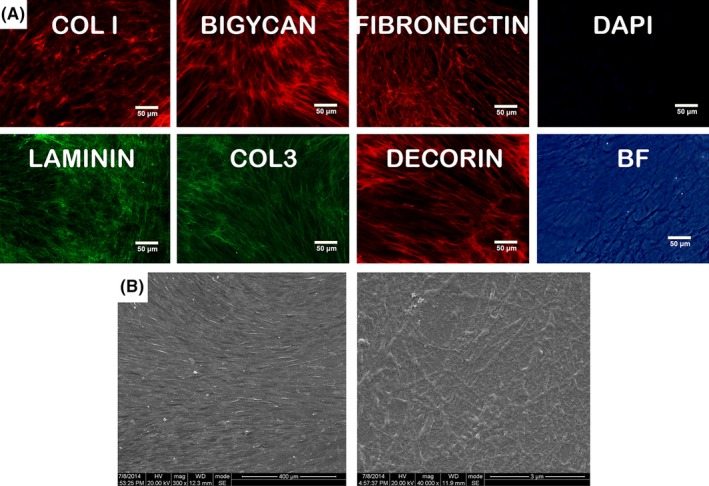
Fabrication of DPM. Immune fluorescence showed the DPM owned net fibrils structure that was positive for Col‐I, Col‐III, laminin, decorin, fibronectin, biglycan, which indicated the decellularization preserved the basic ECM proteins and structure (A). The decellularization was visualized by staining nuclei with the fluorescent dye DAPI (A). SEM image (B) showed the microstructure of DPM under 300× and 40 000× amplification, respectively (n=3)