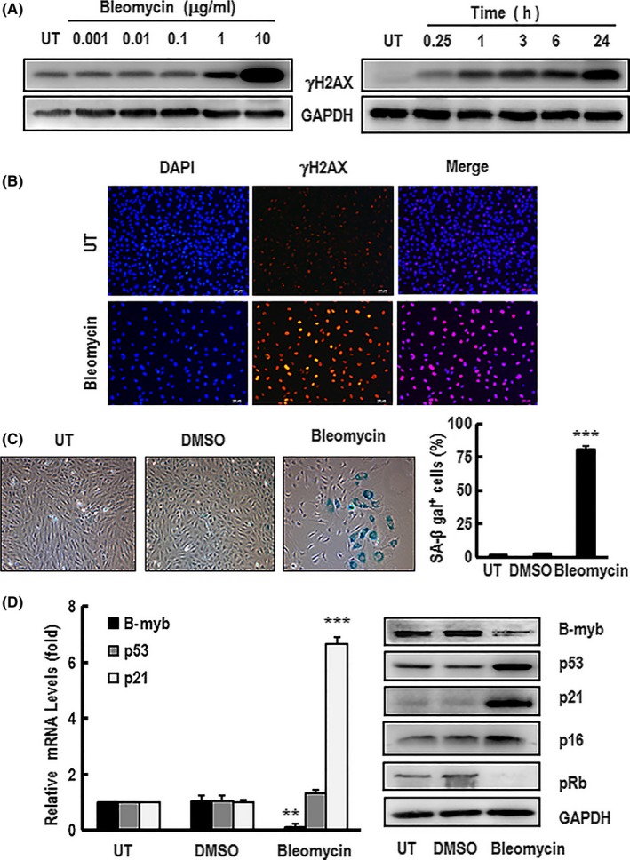
Figure 2.

HAEC premature senescence induced by bleomycin influenced the expression of B‐myb, p53, p21, p16 and pRb. A, The dose‐response curves and time course of activation of γH2AX were detected by western blotting after HAECs were treated with bleomycin in the indicated concentrations for 24 h or 10 μg/mL bleomycin for the time period indicated. GAPDH was used as a loading control. These blots were obtained from one of three independent experiments. B, Cells were stimulated with 10 μg/mL bleomycin for 24 h before immunostaining with antibodies against γH2AX (red) and counterstaining with DAPI (blue). A representative group image of stained cells was observed under fluorescence microscope (×100 magnification). HAECs were treated with 10 μg/mL bleomycin for 60 min following replacement of normal culture medium for 4 d. C, The SA‐β‐gal‐positive cells were observed under inverted microscope (×100 magnification) after the treated cells were stained with senescence‐associated β‐galactosidase (SA‐β‐gal). The percentage rate of SA‐β‐gal‐positive cells was analysed. Data are presented as mean ± SEM of three independent experiments. *** indicates P<.001 compared with the control. D, The expression mRNA and/or protein levels of B‐myb, p53, p21, p16 and pRb in the treated cells were detected by real‐time PCR and western blotting, respectively. GAPDH was used for normalization. The mRNA data are presented as mean ± SEM of three independent experiments. ** and *** indicate P<.01 and P<.001, respectively, compared with the control. A typical group of blots for analysing the expression proteins levels of B‐myb, p53 and p21 in the cells is showed from one of three independent experiments. GAPDH was used as a loading control
