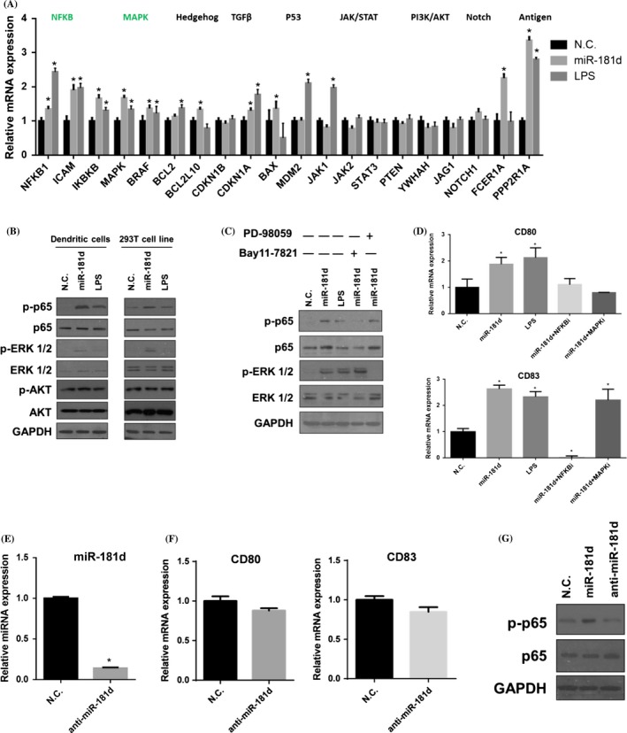
Figure 4.

NF‐κB signalling pathway is modulated by miR‐181d in mDCs (A) qRT‐PCR was performed to examine the expressions of the key components which involved in the signalling pathways. qRT‐PCR measurements were performed in triplicate (n=3). Data represent the means±SEM. (B) Western blotting of p‐p65, p‐ERK 1/2 and p‐Akt in the miR‐181d, LPS and N.C.‐treated dendritic cells (DCs). The same treatment was done in 293T cell line. Data are representative of three independent experiments. (C) DCs were treated with signalling inhibitor PD 98059 and Bay 11‐7821, combined the treatment with miR‐181d transfection, N.C. and LPS. Western blotting showed the p‐p65 and p‐ERK 1/2 expression after the treatment. Data are representative of three independent experiments. (D) CD80 and CD83 mRNA expressions were detected by qRT‐PCR with PD 98059 and Bay 11‐7821, and miR‐181d mimics transfection. qRT‐PCR measurements were performed in triplicate (n=3). (E) DCs were transfected with anti‐miR‐181d (50 nmol/L) for 24 hours on day 6 culture. Expression of miR‐181d was assessed by qRT‐PCR. (F) CD80 and CD83 expression were detected by qRT‐PCR in iDC and anti‐miR‐181d‐transfected iDC. (G) Expression of p‐p65 and p‐65 proteins was detected by Western blotting in DCs transfected with miR‐181d, anti‐miR‐181d or N.C. respectively. GAPDH was used as the internal control. Data represent the means±SEM (Student's t tests; *P<.05)
