
Figure 5.
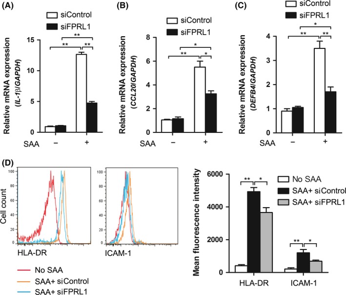
FPRL1 mediates the activation of keratinocytes induced by SAA. The siRNA‐transfected keratinocytes were treated with SAA for 24 h. (A) IL‐1β, (B) CCL20 and (C) DEFB4 gene expressions were then assessed by real‐time PCR and normalised against the amount of GAPDH mRNA. (D) Expression levels of HLA‐DR and ICAM‐1 were analysed by flow cytometry. Representative histograms are shown. The data from (A‐D) represent the mean ± SEM of three experiments (*P<.05, **P<.01, one‐way ANOVA)