
Figure 2.
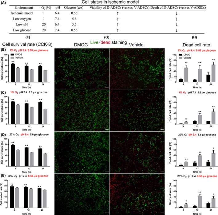
Increased survival of D‐ADSCs in the ischaemic model. After being seeded in 96‐well plates, 1×10⁴ D‐ADSCs (100 μmol/L dimethyloxalylglycine for 4 days) or V‐ADSCs were placed in four different environments (A), at 6, 12 and 24 hours, and CCK‐8 (F) and Live (green)/dead (red) assay (fluorescence (G), cell death rate (H)) were performed. The results display increased survivability of D‐ADSCs not only in the ischaemic model (B) characterized by low oxygen (1% O2), low glucose concentration (0.56 μmol/L) and low pH value (6.4) but also in each of these unfavourable environments individually (C, D, and E). **P<.05, **P<.01, scale bar=200 μm. ADSC, adipose‐derived stem cell