Figure 6.

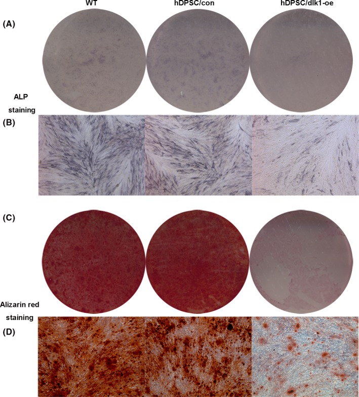
ALP staining and mineralized nodule formation results. (A and B) A significantly lower ALP staining in the hDPSC/dlk1‐oe group on 7 d compared with in the hDPSC/wt and hDPSC/control groups was found. (C and D) The amount of mineralized nodules formed in the hDPSC/dlk1‐oe group was much less than control groups (A, C: scanner; B and D: phase‐contrast microscopy, 50×)
