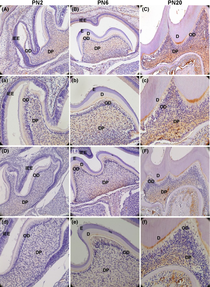
Figure 8.

p‐ERK1/2 and NOTCH1 protein expression in the first maxillary molar in mice were detected by immunohistochemical analysis. A(a), B(b) and C(c) show the result of p‐ERK1/2. D(d), E(e) and F(f) show the result of NOTCH1. p‐ERK1/2 expression pattern was much more similar with the DLK1 than NOTCH1 during the tooth development. (IEE: inner enamel epithelium; OD: odontoblast; DP: dental pulp; E: enamel; D: dentin; A, B, C, D, E and F: 200×; a, b, c, d, e and f:400×)
