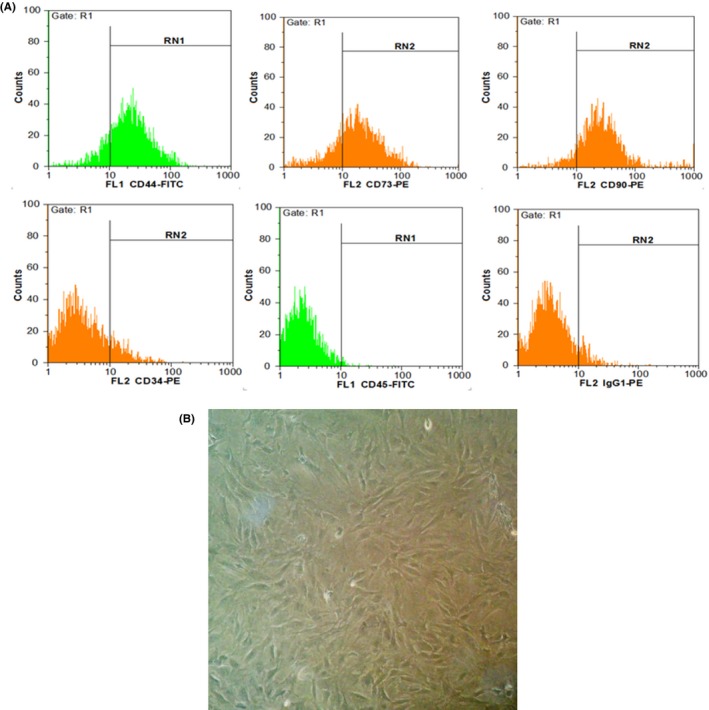
Figure 1.

Characterization of MSC using flow cytometry. A, MSCs were positive for CD73, CD44 and CD90, while they were negative for CD34 and CD45 (hematopoietic markers). B, MSCs after fourth passage in 90% confluency. Cells showed fibroblast like morphology (long and thin) under phase contrast microscope
