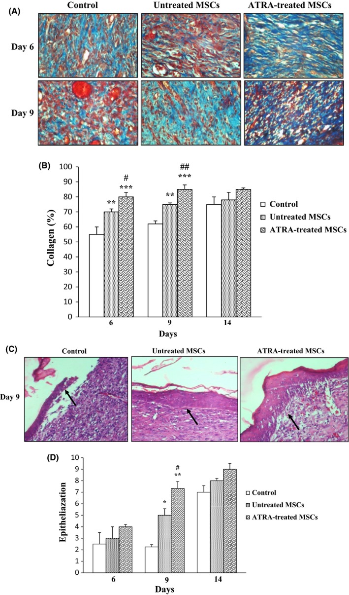
Figure 8.

Effect of ATRA on collagenization and epithelialization. A, Masson's trichrome staining was used for assessment of collagenization. Fibers of collagen are stained blue and arrows signify collagen fibers. Wounds injected with ATRA‐treated MSCs and untreated MSCs had higher tissue collagenization compared to those injected with PBS at 6 and 9 days. Intensity of staining was compared with normal skin on day 0 was determined by NIH image J. B, Comparison between percent of collagen fibers in groups within 14 days. C, Epithelialization was evaluated using H&E staining of wound sections. Arrows signify epithelium. Wounds injected with ATRA‐treated MSCs had higher tissue epithelialization compared to untreated MSCs and PBS groups at 9 days. D, Comparison between the number of epithelium layers in groups. Data are represented as mean±SEM. (n=30) *P<.05, **P<.01, ***P<.001 vs control group and # P<.05, ## P<.01 vs untreated MSCs
