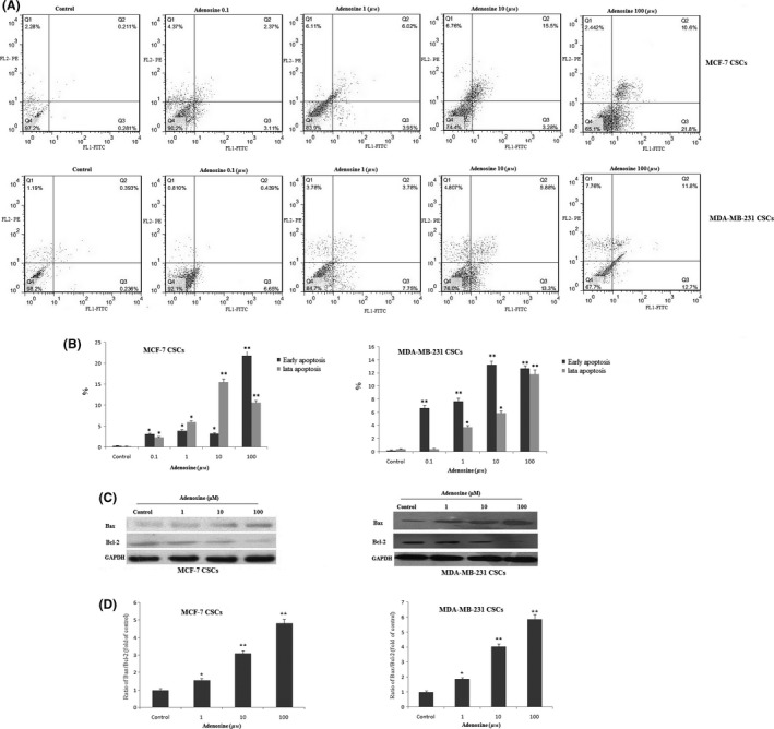
Figure 5.

Detection of apoptosis in breast cancer stem cells (CSCs). Flow cytometric analysis of breast CSCs after treatment with adenosine (A). After treatment with adenosine apoptosis gradually increased in breast CSCs (B). The expression levels of Bax and BCL‐2 proteins were determined by Western blot (C). Quantification analysis of Bax/Bcl‐2 protein expression ratio (D). The results shown represent the mean ± SD of three independent experiments. *P<.05; **P<.01 compared with the control group
