
Figure 1.
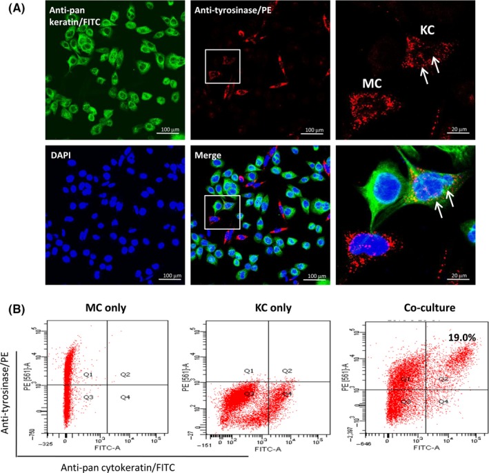
Quantitative assessments for melanosome transfer in MC‐KC co‐culture. (A) Human primary MCs and primary KCs were co‐cultured on coverslips at a ratio of 1:10 for 24 h. The co‐cultured cells were incubated with the mixture of two primary antibodies, including anti‐tyrosinase antibody conjugated with PE and anti‐pan cytokeratin antibody conjugated with FITC. Cell nuclei were then stained using DAPI solution. Higher magnifications of the boxed areas in the middle panel are shown on the right, white arrows indicate the representative image of melanosome transfer that the tyrosinase‐positive particles (red) are existed in cytokeratins‐positive cells (green). Scale bar: 100 μm. (B) Flow cytometry assay was performed to quantitatively analyse melanosome transfer rate using the same antibodies that were used for immunofluorescent double‐staining. The number of melanosome‐containing KCs was shown in the upper right quadrant. MCs only (in the upper left quadrant) and KCs only (in the lower right quadrant) were used as controls