
Figure 2.
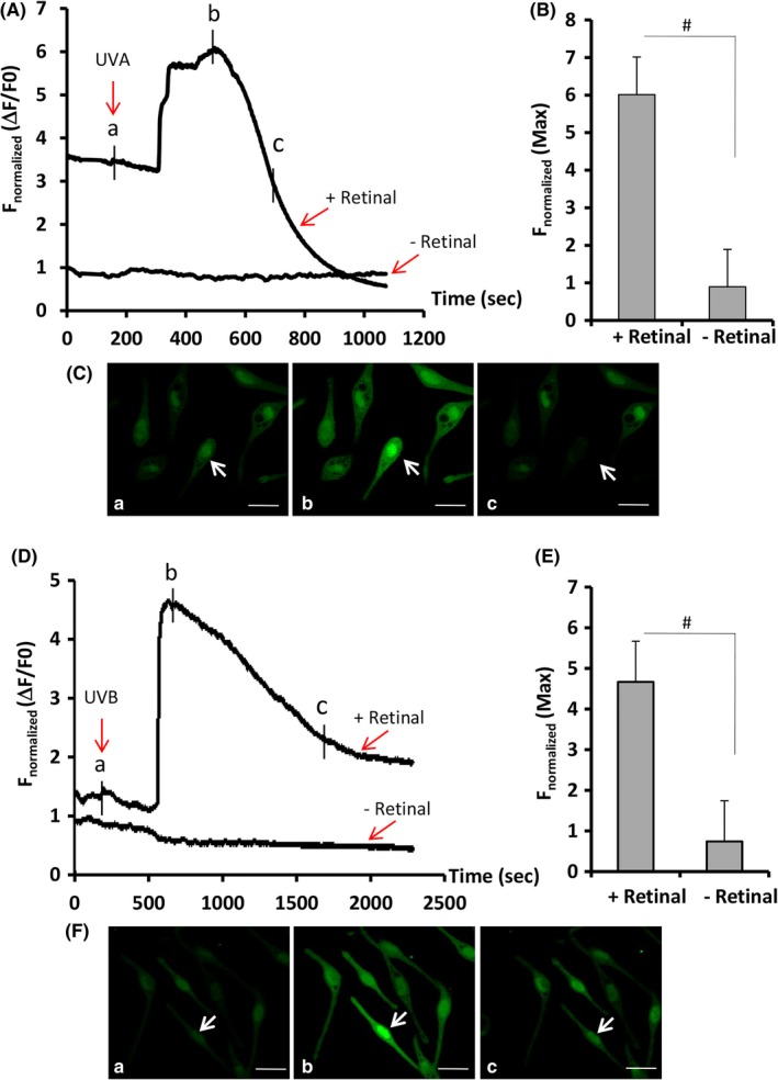
Ca2+ influx profiles of human MCs in response to ultraviolet A (UVA) or ultraviolet B (UVB). Human primary MCs grown in chamber slides were loaded with 2 μM Fluo‐4 AM dye, and cytosolic Ca2+ was monitored using a two‐photon confocal microscopy as described in the Materials and Methods. The fluorescence intensity of Fluo 4‐loaded MCs in response to 3 J/cm2 UVA (A‐C) or 20 mJ/cm2 UVB (D‐F) was recorded in the presence or absence of 10 μM all‐trans retinal. After exposure to UVA or UVB, the images of intracellular Ca2+ changes in representative cells (indicated by a white arrow) captured at a time in A and D (denoted by a, b, and c) are shown in the bottom panels (C and F). Changes of fluorescence intensity were calculated and normalized for 10 cells in each experiment (A and D). Bar graphs show the mean of normalized maximum fluorescence intensities from three independent experiments with at least 10 cells counted per experiment. # P < .01