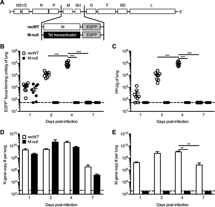
Figure 1. No infectious progeny is detected following RSV M-null infection in vivo.
(A) Diagram depicting the genome content of RSV recWT and RSV M-null. (B-E) BALB/c mice were infected with either RSV recWT or RSV M-null, and lungs were harvested on days 1, 2, 4, and 7 p.i. (B) EGFP+ focus-forming units and (C) infectious PFU were quantified in the lung by plaque assay using H2-M helper cells. Open (recWT) and closed (M-null) circles represent values for individual mice and lines indicate mean ± SEM of 2 independent experiments (n=8). (D) RSV N gene and (E) RSV M gene copy numbers per lung were determined by real-time PCR. Data are presented as mean ± SEM of representative results from 1 of 2 independent experiments (n=4). Dashed lines denote the limit of detection for each assay. Groups were compared using one-way ANOVA, ** p<0.01, *** p<0.001.

