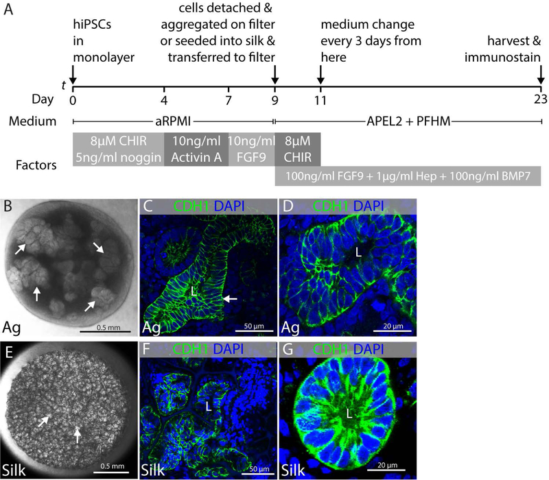
FIGURE 3.
Epithelial differentiation of kidney progenitors. Experiments were performed in triplicate and data representative of a minimum of 3 independent experiments are shown. (A) Schematic illustration of protocol showing directed differentiation of hiPSCs into kidney progenitors on day 9 and subsequently into epithelial structures after making aggregates and seeding in silk scaffold. (B–D) Epithelial structures that differentiate in aggregates of cells derived from directed differentiation cultured in organotypic conditions. (B) Representative stereomicroscope image of kidney organoid generated from hiPSCs at air-liquid interface showing presumptive epithelial clusters (arrows). (C) Representative immunofluorescent image showing CDH1+ tubular epithelial structure with lumen (L) in aggregated cells from directed differentiation (arrow). (D) High magnification micrograph of epithelial tubule with lumen in aggregated cells from directed differentiation. (E–G) Epithelial structures that differentiate in cells from directed differentiation that have been seeded into silk scaffolds. (E) Representative stereomicroscope image of silk scaffold seeded with cells derived from directed differentiation showing clusters of cells within the pockets of the silk scaffold (arrows). (F) Representative immunofluorescent image showing CDH1+ epithelial tubules with lumens (L) within the pockets of the silk scaffold. (G) High magnification micrograph of CDH1+ epithelial tubule with lumen (L) within a pocket of the silk scaffold.

