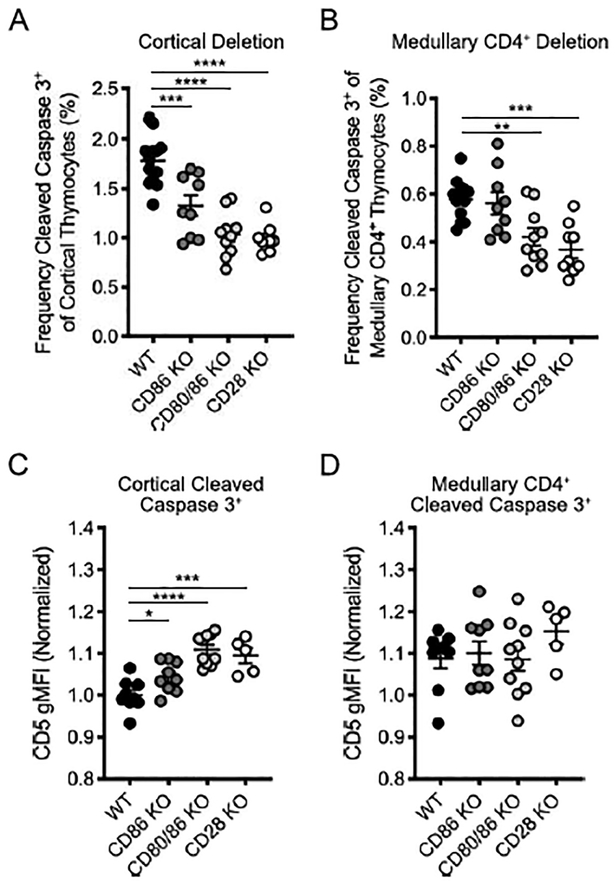
Figure 4. Co-stimulatory molecules CD80 and CD86 are required for both cortical and medullary clonal deletion.
(A) Frequency of cleaved caspase 3+ cells among CD5+TCRβ+ cortical (CCR7−) thymocytes (as gated in Figure 1A) in WT, CD86 KO, CD80/86 KO, and CD28 KO mice. (B) Frequency of cleaved caspase 3+ cells among CD5+TCRβ+ medullary (CCR7+) CD4+ thymocytes (as gated in Figure 1A). (C) CD5 gMFI of cortical deleted thymocytes (normalized to mean of WT CD5 gMFI) (from A). (D) CD5 gMFI of medullary deleted CD4+ thymocytes (normalized to mean of WT CD5 gMFI) (from B). Each symbol (A, B, C, D) represents an individual mouse. Six- to twelve-week-old male and female mice were used. Small horizontal lines indicate the mean and error bars represent SEM. *P< 0.05, **P< 0.01, ***P< 0.001, ****P< 0.0001. Statistical significance was determined by ordinary one-way ANOVA with Holm-Sidak’s multiple comparisons test (A, B, C, D). Data are pooled from at least three independent experiments (A, B, C, D).

