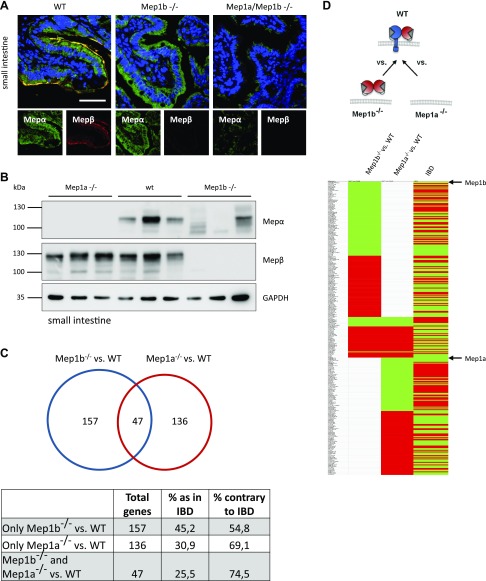
Figure 7.
Expression and transcriptional analysis of meprin heterodimer in vivo. A) Immunofluorescence microscopy of small intestine from WT, Mep1b−/−, and Mep1a−/− mice using specific meprin antibodies. Staining revealed absence of meprin α at the cell surface in Mep1b−/− mice. Scale bar, 40 µm. B) Immunoblotting of small intestine lysates of WT, Mep1b−/−, and Mep1a−/− mice using specific meprin antibodies. GAPDH served as loading control. C) Analysis of RNA sequencing of WT, Mep1b−/−, and Mep1a−/− mice small intestine samples. Obtained data were compared with transcriptome analyses of IBD patients (32), and matching and overlapping genes of each condition were depicted. D) Representation of matching up- and down-regulated genes in WT, Mep1b−/−, and Mep1a−/− mice small intestine samples and IBD patients. Green, down-regulation, red, up-regulation. Mep, meprin.

