Figure 7.
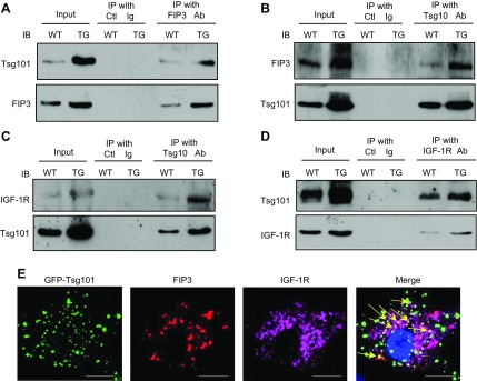
Tsg101 interacts with FIP3 and IGF-1R. A, B) Coimmunoprecipitations using anti-FIP3 (A) or anti-Tsg101 (B) as primary antibody, showing that the Tsg101 and FIP3 association was enhanced in TG (line G) hearts. C, D) Coimmunoprecipitations, using anti-Tsg101 (C) or anti–IGF-1R (D) as primary antibody, demonstrated that the Tsg101 and IGF-1R association was enhanced in TG (line G) hearts. WT and TG heart homogenates were used as input. E) Representative images showing partial colocalization for FIP3 and IGF-1R in Ad.Tsg101-infected NRCMs (yellow arrows). Similar results were observed in 3 additional, independent experiments. Ctl, control; IB, immunoblot; IP, immunoprecipitates. Scale bars, 20 μm.

