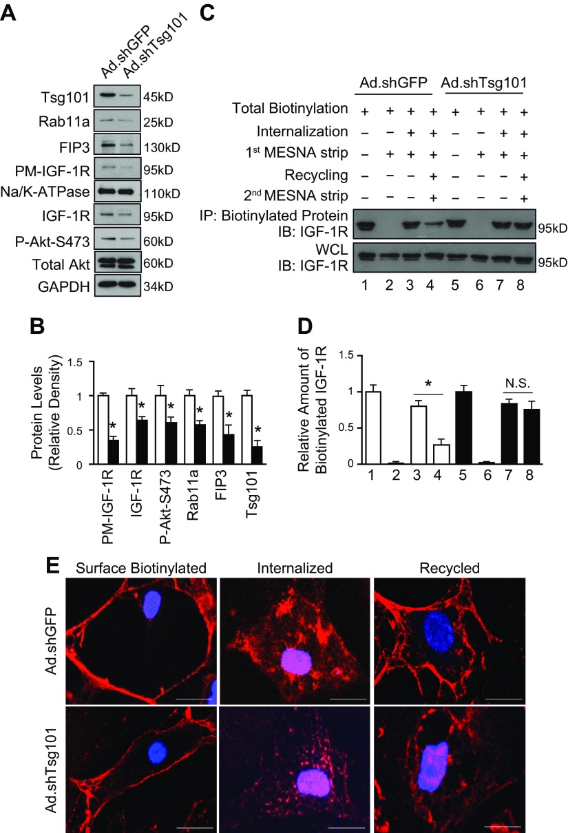
Figure 9.
KD of Tsg101 in neonatal myocytes blocks recycling of IGF-1R. A, B) Representative immunoblots (A) and quantitative analyses (B) showing protein levels of PM IGF-1R, total IGF-1R, Akt phosphorylation, Rab11a, FIP3, and Tsg101 in Ad.shTsg101 and Ad.shGFP NRCMs. GAPDH was loading control for total protein, total Akt was loading control for Akt phosphorylation, and Na/K-ATPase was loading control for PM; n = 4 for independent experiments; *P < 0.05 vs. Ad.shGFP cells. C, D) Representative immunoblots (C) and quantitative analyses (D) of streptavidin IPs visualized using an IGF-1R antibody. WCLs were used as loading controls. Four independent experiments were performed for recycling assays. KD of Tsg101 inhibited recycling of IGF-1R, whereas recycling of IGF-1R was uninhibited in Ad.shGFP cells. E) Representative images of immunofluorescence staining with streptavidin Alexa Fluor 594 conjugate after IGF-1R biotin was internalized and recycled in Ad.shTsg101 and Ad.shGFP NRCMs. IP, immunoprecipitates; N.S., not significant; PM, plasma membrane protein; WCL, whole-cell lysate. n = 4 plates, 25–30 cells per plate. Scale bars, 5 μm. *P < 0.001.

