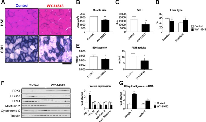
Figure 5.
WY-14643 administration causes muscle atrophy and mitochondrial alterations. A) Representative images of hematoxylin and eosin (H&E)– and SDH-stained 10 μm-thick sections of tibialis anterior muscle excised from control and WY-14643–treated mice. Scale bar, 100 μm. B) CSA in tibialis anterior muscle sections from mice fed a diet enriched with WY-14643. C) Quantification of SDH integrated density in control and WY-14643–treated mice. Data are expressed as arbitrary units (a.u.). D) Number of oxidative (dark blue) and glycolytic (light blue) fibers (expressed as percentage of control). E) SDH and PDH enzymatic activities in the gastrocnemius muscle of control and WY-14643–treated animals. Data are expressed as mU/μl for SDH or mU/ml for PDH. F) Representative Western blotting for PDK4, PGC1α, OPA1, Mitofusin 2, and cytochrome c in whole-muscle protein extracts from mice fed control or WY-14643 diets. Tubulin was used as loading control. G) Gene expression levels for atrogin-1 and MuRF1 ubiquitin ligases performed by real-time quantitative PCR. Gene expression was normalized to TATA-binding protein levels. Data (means ± sd) are expressed as fold change vs. control; n = 8. *P < 0.05, **P < 0.01, ***P < 0.001.

