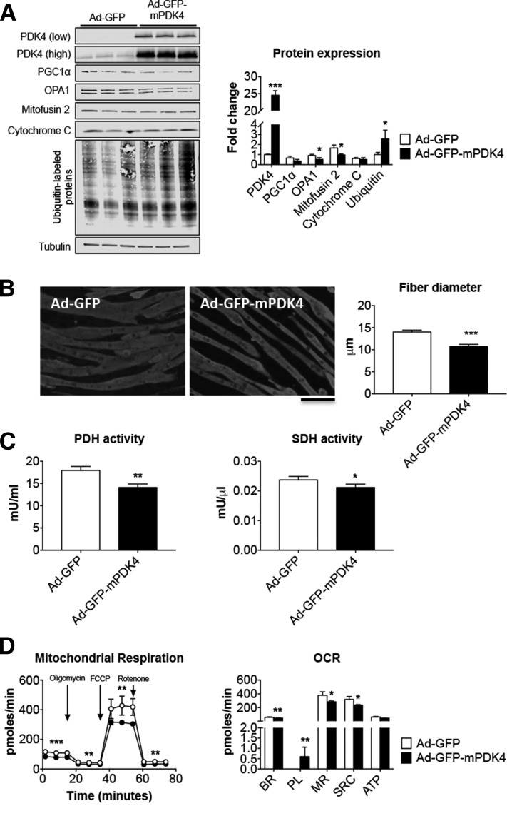
Figure 6.
PDK4 overexpression affects C2C12 myotube size and metabolism. A) Representative Western blotting and quantification for PDK4 (shown with both low and high exposure times), PGC1α, OPA1, Mitofusin 2, cytochrome c, and ubiquitin-labeled proteins in C2C12 myotubes infected with Ad-GFP or Ad-GFP-mPDK4 (n = 3). Tubulin was used as loading control. B) Myosin heavy chain (MyHC) immunofluorescent staining in C2C12 myotubes infected with Ad-GFP or Ad-GFP-mPDK4 (n = 3) and myotube size quantification. On average, 250–350 myotubes were measured per experimental condition, in triplicate. Scale bar, 100 μm. C) Pyruvate dehydrogenase (PDH) and succinate dehydrogenase (SDH) enzymatic activities in C2C12 myotubes infected with Ad-GFP and Ad-GFP-mPDK4. Data are expressed as milliunits per millilter for PDH, or milliunits per microliter for SDH. D) Mitochondrial respiration and oxygen consumption rate (OCR) in C2C12 myotubes infected with Ad-GFP or Ad-mPDK4 (n = 3). ATP, adenosine triphosphate; BR, basal respiration; MR, maximal respiration; PL, proton leak; SRC, spare respiratory capacity. Data (means ± sd) were expressed in picomoles per minute. Data (means ± sd) are reported as fold change vs. Ad-GFP. *P < 0.05, **P <0.01, ***P < 0.001 vs. Ad-GFP (significance of the differences).

