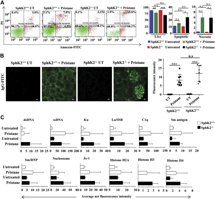
Figure 4.
Absence of SphK2 enhances pristane-induced apoptosis of peritoneal cells. A) Peritoneal-lavage cells from SphK2+/+ to SphK2−/− mice untreated or injected with pristane were stained with PI and annexin V–FITC and analyzed by FACS. Representative FACS analyses are shown in the left panels. The right panel shows average populations of live cells (PI and annexin negative), necrotic cells (PI positive and annexin negative), and early and late apoptotic cells (annexin positive). Data are expressed as means ± sem (n = 7 mice each in SphK2+/+ groups, and n = 6 mice each in SphK2−/− groups). Statistical significance was determined by ANOVA with a post hoc Tukey’s test. B) Glomerulonephritis in renal sections were determined by deposition of IgG-FITC immunofluorescence after staining with goat anti-mouse IgG antibody. Scale bars, 10 µm. Data from 8 fields were quantified by ImageJ software. Statistical significance was determined by ANOVA with a post hoc Tukey’s test. C) IgG autoantibodies in serum from SphK2+/+ to SphK2−/− mice untreated or injected with pristane were measured fluorometrically with an autoantigen microarray as described in Materials and Methods. Data are presented as average net fluorescence intensities; n.s., not significant. *P < 0.05, **P < 0.005, ***P < 0.001.

