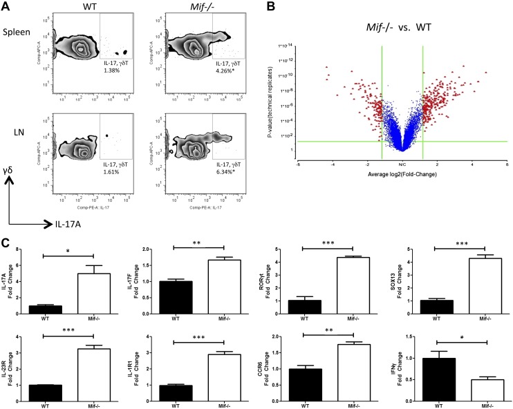
Figure 4.
Higher production of IL-17A in γδ T cells of Mif−/− mice in response to the Mycobacterium glycolipid, LM. Spleen and lymph node (LN) cells of WT or Mif−/− C57BL/6 mice were stimulated in vitro with 1 µg/ml LM. A) After 72 h of culture, IL-17A expression was determined by flow cytometry. For microarray and real-time PCR analysis, the γδ T cells were separated by magnetic cell sorting after 18 h of culture. B) The microarray results are shown as a volcano plot. C) The levels of IL-17A, IL-17F, RORγt, SOX13, IL-23R, IL-1R1, CCR6, and IFN-γ mRNA were examined by qPCR. APC, allophycocyanin; comp, compensation; PE, phycoerythrin. Error bars denote sd. All data shown are representative of 3 replicates. *P < 0.05, **P < 0.01, ***P < 0.001 (unpaired Student’s t test).

