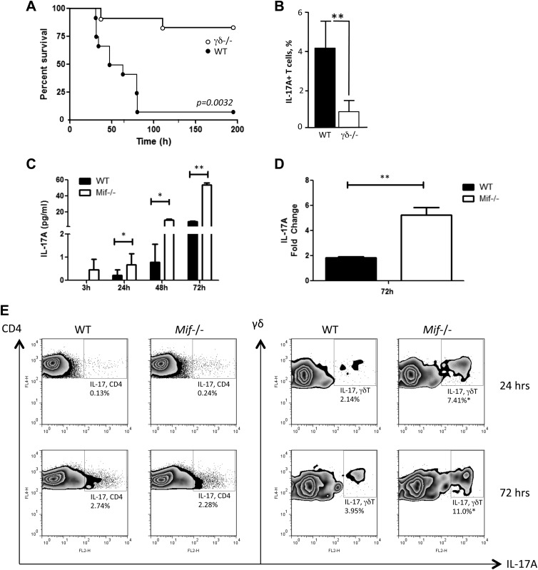
Figure 5.
Production of IL-17A in response to M. bovis infection. A) Survival of WT and γδ TCR−/− (γδ−/−) C57BL/6 mice infected intraperitoneally with M. bovis (n = 9 mice/group, Gehan-Breslow-Wilcoxon test. P = 0.0032). B) Flow cytometry analysis of peritoneal lavage fluid content of total IL-17A+–expressing T cells as percent of total recorded cells (n = 4 samples per group). C–E) Spleen cells of WT or Mif−/− C57BL/6 mice were infected with M. bovis in vitro at a multiplicity of infection of 50. At indicated time points, the IL-17A level in the supernatants was measured by ELISA (C), and the cellular IL-17 mRNA level was examined by real-time PCR (D). Production of IL-17A by CD4 αβ T cells and γδ T cells was determined by flow cytometry (E). FL4-H, flourescence 4 channel height Error bars denote sd. *P < 0.05, **P < 0.01 (unpaired Student’s t test). All data shown are representative of replicate experiments.

