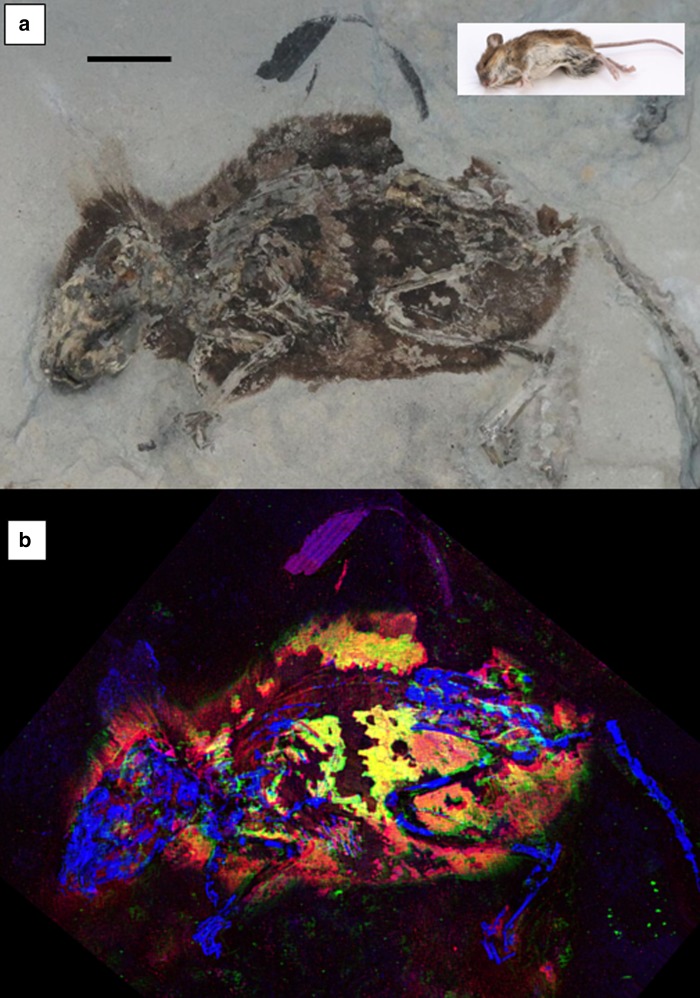
Fig. 1.
Optical and X-ray images of Apodemus atavus “lateral” fossil. a Optical image of “lateral” fossil A. atavus (GZG.W.20027b) with the inset of extant A. sylvaticus in the upper right for comparison (scale bars = 1 cm). b False-color SRS-XRF image reveals exceptional preservation of integument as well as bone. This image is a combination of three maps, two standard single-element maps (blue = P, green = Zn), plus a third map which has been produced to especially emphasize the distribution of a specific oxidation state of organic sulfur (red = S in thiol) in order to highlight the clear correlation between the distribution of Zn and organic sulfur which together appear as bright yellow. (Optical photograph by P.L.M.)

