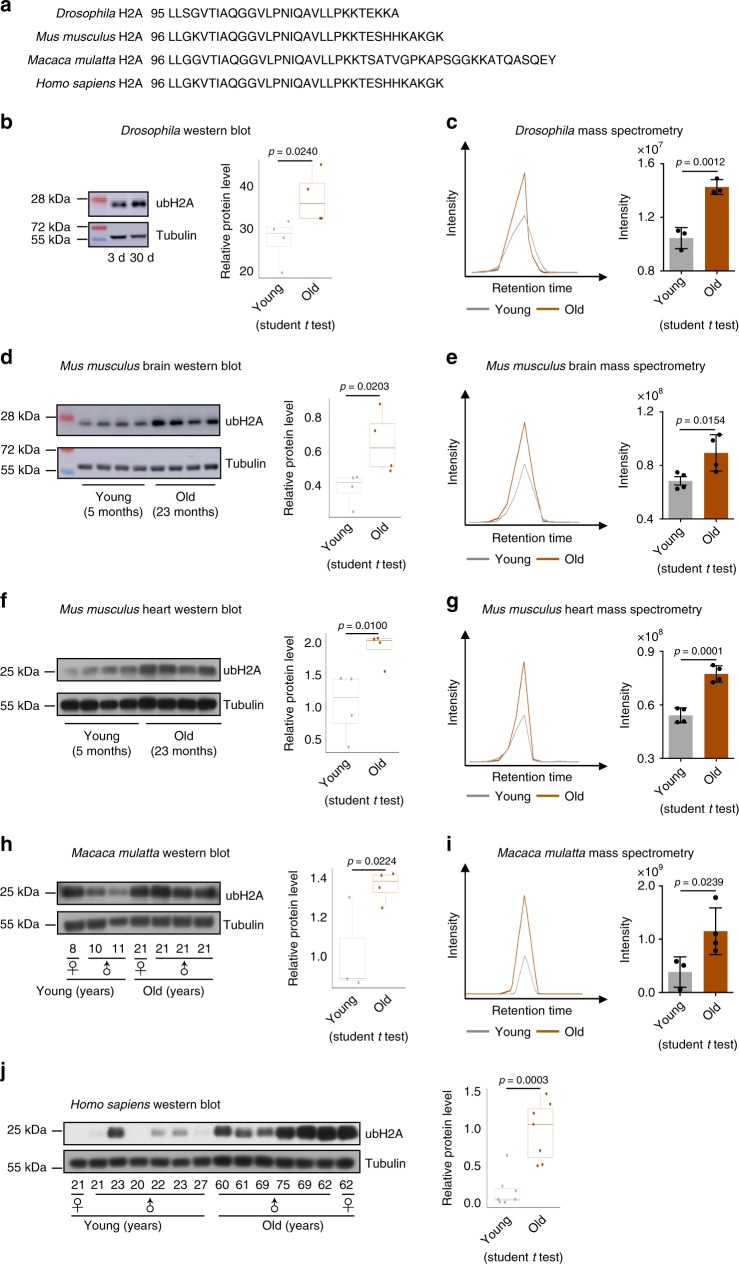
Fig. 3.
H2A mono-ubiquitylation increases with age in Drosophila, mouse, monkey, and human. a Alignment of amino acid sequence showing striking conservation of H2A in Drosophila, mouse, monkey, and human. b, d, f, h, j Western blotting (left) and quantification (right) show an evolutionarily conserved increase of ubH2A with age. Western blotting was from head tissues of 3d and 30d old male flies (n = 4 independent biological repeats) (b), brain and heart tissues of 5 months (n = 4) and 23 months (n = 4) old male mice (d, f), parietal lobes of young (n = 3) and old (n = 4) rhesus macaque (h), and prefrontal cortex of brain tissues of young (n = 7) and old (n = 7) male or female humans (j). The median values are marked at the center. The boxes extend from the 25th to 75th percentiles. The p-values are calculated by using the student t test. c, e, g, i MS quantifications confirm the increase of ubH2A with age in Drosophila (n = 3 independent biological repeats)(c), mouse brain (n = 4 for both young and old groups) (e), mouse heart (n = 4 for both young and old groups) (g), and monkey (n = 3 for young, n = 4 for old) (i). The ubH2A was measured by PRM-based targeted mass spectrometry method. Representative XICs of the y3 ion were shown. The quantification was done by using the summed intensities of y3, y4, y5, y6, and y7 ions. Data are shown as mean ± s.d. The p-values are calculated by using the student t test

