Figure 1.
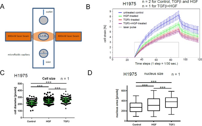
Effects of TGFβ- and HGF-stimulation on rigidity and morphology of NSCLC cells. (A) Working principle of the cell optical stretcher: uniaxial cell elongation from the inital diameter L to L′ = L + dL under the impact of optical forces. (B) Creep-and-recovery curves of untreated, HGF-, TGFβ-treated and co-stimulated H1975 cells. Solid lines show the mean cell strain of bootstrap sample means. Error bars indicate 95% confidence interval which is given by two-fold standard deviation of bootstrap sample means. Cells were treated with 2 ng/ml TGFβ, or 80 ng/ml HGF or combination of both for 24 h in growth factor-depleted medium. Trypsinized cells were injected into the microfluidic system of cell optical stretcher. At least 300 cells per condition were measured. (C) Growth factor treatment leads to the increase of cell size of H1975 cells. Cells were treated with 2 ng/ml TGFβ, or 80 ng/ml HGF or left untreated for 24 h in growth factor-depleted medium, trypsinized and measured on cell optical stretcher. Cell diameter prior to laser-induced cell stretching was measured and compared between the conditions. (D) H1975 cells were seeded on 24-well plate, treated with 2 ng/ml TGFβ, or 80 ng/ml HGF or left untreated for 24 h in growth factor-depleted medium, stained with Hoechst and imaged with a wide-field fluorescence microscope (Olympus). ImageJ was used to quantify the nuclei area. Center lines show the medians; box limits indicate the 25th and 75th percentiles; whiskers extend 1.5 times the interquartile range from the 25th and 75th percentiles. ***p < 0.001 according to Mann–Whitney test. n indicates the number of independent repetitions.

