Figure 3.
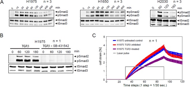
TGFβ/Smad signaling pathway activation in response to TGFβ-stimulation. H1975, H1650 and H2030 cells were kept in serum-free medium overnight. Cells were stimulated with 2 ng/ml TGFβ and lysed at the indicated time points. Subsequently, Smad2/3 immunoprecipitation was followed by immunoblotting. (A) Immunoblotting data of H1975, H1650 and H2030 cells. One representative example is shown. (B) Treatment with TGFβR inhibitor SB-431542 (10 μM) completely abolishes Smad2/3 phosphorylation upon TGFβ-treatment. (C) TGFβ-induced reduction of cell deformability is abolished upon application of TGFβR inhibitor. H1975 cells were treated with 2 ng/ml TGFβ, in presence or absence of 10 μM SB-431542 inhibitor for 24 h. Trypsinized cells were injected into the microfluidic system of cell optical stretcher. At least 300 cells per condition were measured. n indicates number of independent repetitions.

