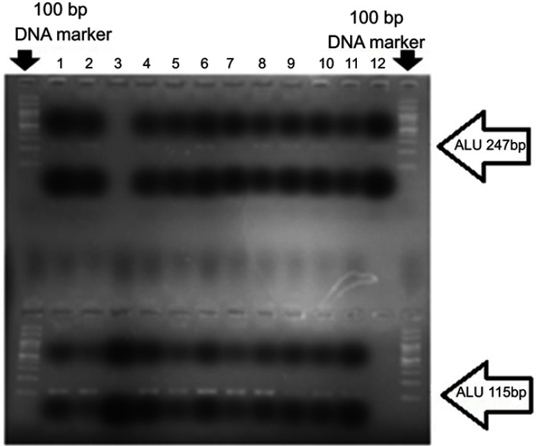
Figure 1.
A number of cancerous samples were examined after amplification by real-time PCR. The products were mixed with loading buffer and loaded on 2% agarose gel. Lanes 1–12 are examined samples. The sequences of ALU247 (above) and ALU115 (at the bottom) are clearly visible. The first and the last wells show the 100 bp DNA marker.

